![]()
Testing Guides
Want to take a deeper dive into specific test types that are performed on Grason-Stadler equipment? The following testing guides will give a general overview of what the test is, where it can be applied, how to prepare for testing, what are the required instructions for the patient, and provide additional information about how to perform the test on an appropriate GSI device. For more information about testing on your GSI device, make sure to read the user manual. Additionally, the video library has some quick tutorials on basic device operation for a number of GSI screening products. Check out our testing guide for each of the testing categories below!
Exposure to high levels of sound - unmuffled lawn mowers, many power tools, motorcycles, rock music, gunfire - tends to create a temporary threshold shift (TTS) which diminishes with time after exposure.
Any patient tested soon after such exposure will manifest a hearing loss that does not accurately reflect the normal hearing threshold. It is important that the testing procedure prescribe some time interval, usually 16 hours, between the high level sound exposure and the actual hearing test.
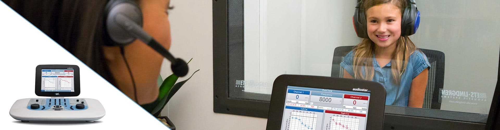
Speech audiometry is an important component of a comprehensive hearing evaluation. There are several kinds of speech audiometry, but the most common uses are to:
- Verify the pure tone thresholds
- Determine speech understanding
- Determine most comfortable and uncomfortable listening levels
The results are used with the other tests to develop a diagnosis and treatment plan.
Special test overviews include:
ABLB
SISI
Stenger
Masking
Pediatric Noise
The Audible Contrast Threshold (ACT) test is a new clinical test that measures binaural spectro-temporal modulation sensitivity to quickly quantify a person’s real-world ability to understand speech-in-noise.
Often, the primary complaint of hearing-impaired people is that they experience difficulty understanding others in background noise. Speech in Noise Audiometry testing is used to help diagnose this and understand the severity of hearing loss and the ability of the patient to hear in background noise. One of these tests is the Quick Speech in Noise Test (QuickSIN).
The BKB-SIN test is a speech-in-noise test developed for children and adult cochlear implant users. It includes normative data for adults, adults with cochlear implants, and children in various age ranges. The BKB-SIN Test is a flexible tool that can be applied clinically in a variety of ways.
Tone decay is a legacy test on the GSI AudioStar Pro and the GSI Pello that evaluates abnormal adaptation to sound caused by a retrocochlear pathology, such as an acoustic neuroma. This is a subjective evaluation that can add diagnostic value to an audiometric evaluation.
The TEN Test is a clinical test that is used to identify dead regions of the cochlea. If a cochlear dead region is identified, the results can be used in the hearing aid fitting by making adjustments to the hearing aid gain. TEN test results may also be helpful when counselling the patient and cochlear implant candidacy.
Tympanometry provides an objective means for determining the amount of mobility present within the eardrum and the ossicular chain. It is, however, important to keep in mind the fact that the amount of mobility present within the ossicular chain may be camouflaged by a scarred or thickened eardrum. Acoustic energy, commonly referred to as the probe tone (226 Hz, 678 Hz or 1000 Hz) is introduced into a hermetically sealed ear canal by means of a loudspeaker located within the probe box. The intensity of this tone is monitored via a microphone, also located within the probe box. Measurements are taken at fixed time intervals.
WideBand Tympanometry is a specialized test designed to evaluate middle ear function across a wide frequency range by utilizing a wideband click stimulus. Unlike traditional tympanometry, WideBand Tympanometry allows for obtaining tympanograms at multiple frequencies with a single pressure sweep.
The acoustic reflex consists of a response by one or more middle ear muscles to suprathreshold acoustic stimulation of the auditory pathway. To elicit an acoustic reflex, an acoustic stimulus (pure-tone, noise, or click), is presented to the ear canal by a probe or earphone. A portion of this stimulus is carried by the ossicular chain to the cochlea. From the cochlea, the 8th nerve carries the information to the brain stem where a determination is made as to whether the stimulus is sufficiently intense to elicit a response. When a response is elicited, the 7th nerve carries the command to the stapedius muscle to contract. Contraction of this muscle and/or the tensor tympani stiffens the eardrum and the ossicular chain; thereby, decreasing the ease with which sound enters the auditory pathway. Thus, the end result of an acoustic reflex is a slight decrease in the ability of the eardrum and the ossicular chain to conduct acoustic energy to the cochlea.
One purpose of the Eustachian tube is to equalize pressure between the middle ear space and ambient pressure. Normally, the Eustachian tube temporarily opens during a swallow or yawn; thereby, allowing an exchange of air between the middle ear and the nasopharynx. Between swallows, slight fluctuations may occur in the pressure level within the middle ear since the cells which line the middle ear absorb air from the cavity. If the Eustachian tube should remain closed for an extended period of time, a negative pressure (relative to atmospheric pressure) may develop within the middle-ear. This causes the tympanic membrane to retract inward, thus stiffening the eardrum. Air pressure is decreased at the rate of 50 mm H2O/hour if the tube remains closed. In time, fluid may develop within the middle ear space further stiffening the middle ear system and reducing the ability of the ossicular chain to conduct sound to the cochlea. Since a malfunctioning Eustachian tube can lead to middle ear disease and hearing loss, it is helpful to be able to determine the patency of the Eustachian tube in patients who are susceptible to middle ear problems.
Tympanometry performed with a probe tone frequency of 226 Hz provides useful test mode clinical information regarding disorders of the tympanic membrane and the Eustachian tube. The middle-ear system is mainly stiffness-controlled when tympanometry is performed with low frequency probe tones. Ossicular abnormalities affecting the mass-controlled components cause changes in the transmission characteristics of the tympano-ossicular system which are more easily identified with probe tone frequencies that approximate or exceed the resonance frequency of the ear.
Since the microphone measures the response at the entrance to the ear canal, rather than at the site of its origination (the cochlea), the anatomy along the return path (i.e., the ossicular chain and the tympanic membrane) can influence the amplitude of the response. To identify these effects, measure the status of the middle ear using tympanometry screening whenever possible.
This otoacoustic emission has a very small amplitude and gets mixed in with other biological and environmental sounds present in the ear canal. Since the probe microphone detects all of these sounds, the instrument must use signal averaging techniques to separate the emitted response, OAE (signal), from these other sounds (noise).
Transient Evoked Otoacoustic Emissions, or TEOAEs, are sounds generated by the cochlea in response to auditory stimulation. These emissions can be detected in the ear canal of individuals with normal outer hair cell function.
Auditory Evoked Potentials (AEP) can be used to evaluate the integrity of the auditory system and are used to make inferences about hearing. AEPs encompass a series of neurologic events that travel along the entire length of the auditory pathway, from the cochlea to the auditory cortex. There have been as many as 15 AEPs identified within the first 500 ms post-stimulus onset. In order to identify the neural integrity of the auditory system, it is necessary to consider the size and latency of the response and utilize averaging and stimulus parameters to elicit and isolate the AEP interest.
The Automated Auditory Brainstem Response (AABR) is a test method used in newborn hearing screening devices that measures the inner ear and the brain pathway’s response to sounds. When screening for hearing loss, the device will analyze the responses and automatically provide a Pass or Refer result at the end of the test. This is one of the hearing tests that is performed on infants up to 6 months of age and determines when additional diagnostic testing is recommended.
Short-latency potentials evoked through activation of vestibular receptors using sound or vibration are referred to as vestibular-evoked myogenic potentials (VEMP). VEMPs are generated by modulated electromyographic signals and recorded with surface electrodes. A VEMP recorded from the sternocleidomastoid muscle is commonly referred to as the cervical vestibular-evoked myogenic potentials (cVEMP). A VEMP recorded from the inferior oblique muscle has been termed the ocular vestibular-evoked myogenic potentials (oVEMP). These reflexes appear to originate from the otolith organs and thus complement existing methods of vestibular assessment, which are mainly based upon canal function. VEMPs are used clinically to assess the function of the saccule, utricle, and the inferior and superior portions of the vestibular nerve.
The ECochG is a specialized evoked potential that is a measure of the electrical activity in the cochlea. It is made up of the cochlear microphonic (CM), the summating potential (SP), and the action potential (AP) of the eighth cranial nerve. The AP is the Wave I of the ABR. The origin or source of the ECochG is the cochlea therefore the electrode of choice is one that is placed as close to the cochlea as possible.
The ECochG has been used to assist in the diagnosis of Meniere’s Disease, Auditory Neuropathy, and SSCD (Superior Semicircular Canal Dehiscence). It is also used to monitor the success of SSCD repair surgery. The ECochG may be used to identify Wave I when it is not be detected with traditional ABR surface electrodes.
Auditory Middle Latency Responses (AMLR or MLR) are responses to auditory stimuli that occur between 10 and 75 msec after stimulation. The MLR generators are thought to be along the thalamic auditory pathway and the auditory cortex. The MLR can be used to estimate hearing and may be used in cases where auditory processing disorders or central lesions are suspected.
Late Latency Responses (LLR) are responses to auditory stimuli that occur between 50 and 300 msec after stimulation. The LLR originates from the auditory cortex and can assist in evaluating patients with suspected auditory processing deficits, cortical lesions, and when presented in sound field may be used for hearing verification.